

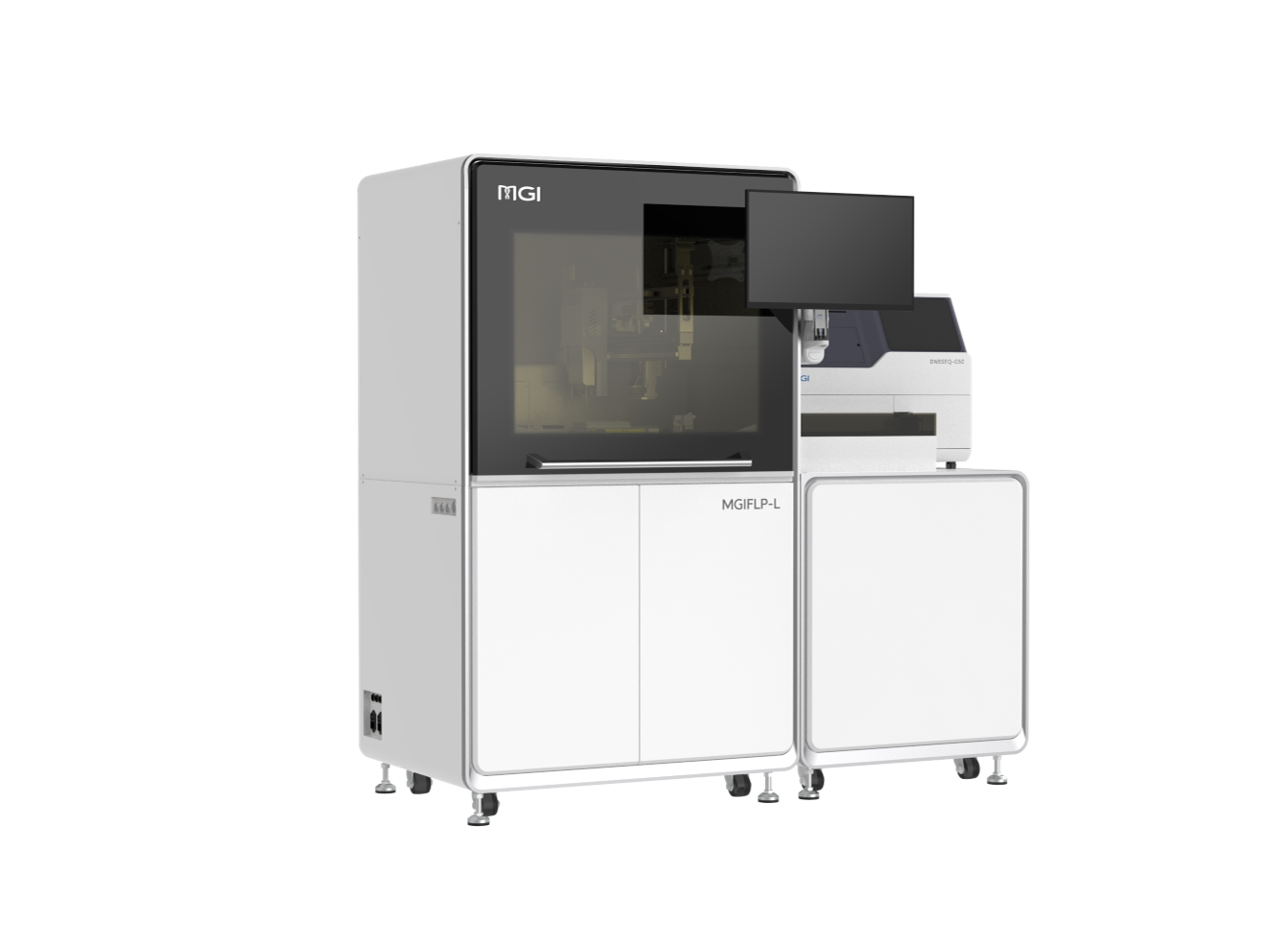
 Sequencer Products: SEQ ALL


 Technologies
 Applications
 Online Resources
 Data Bulletins
 Service & Support
 Introduction
 Newsroom
 Doing Business With Us
 Creative Club

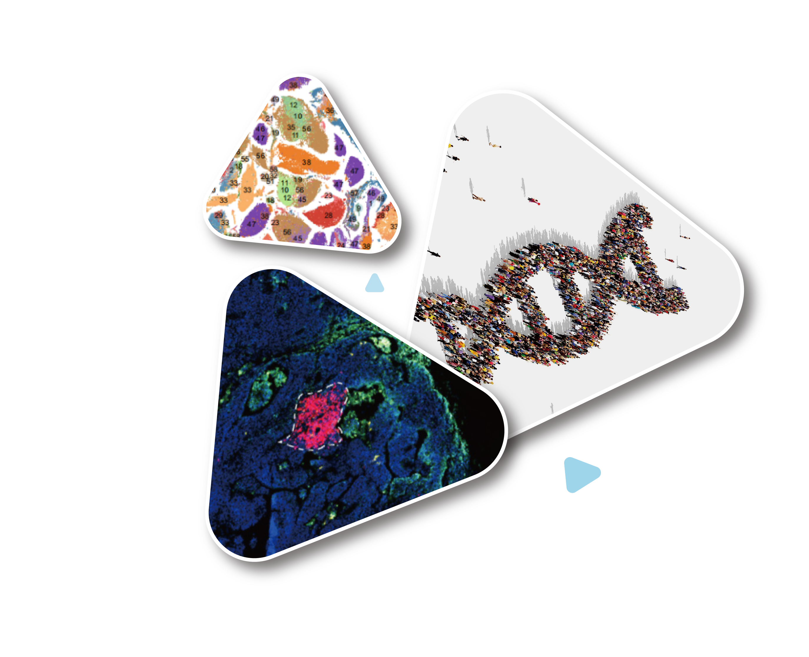
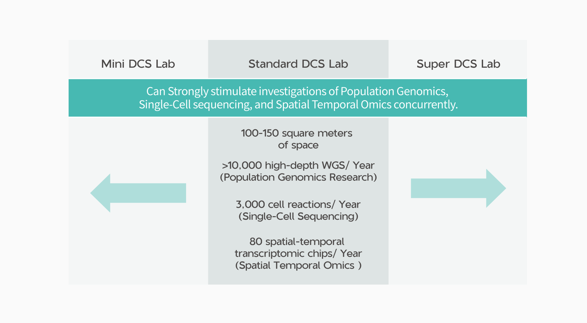
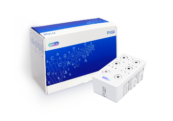
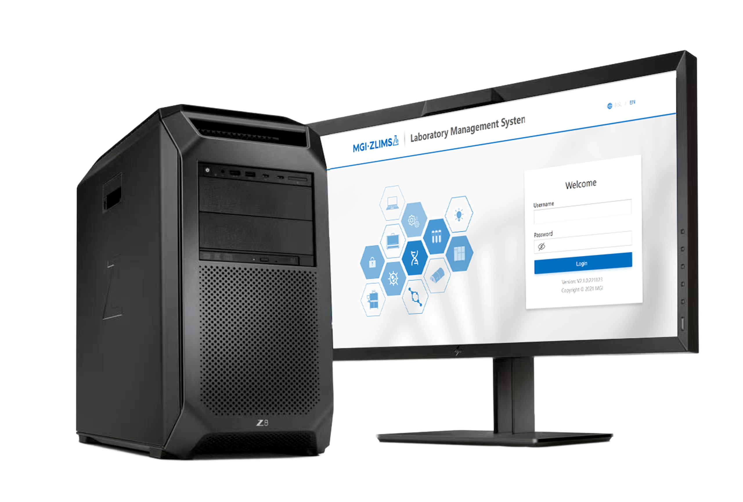
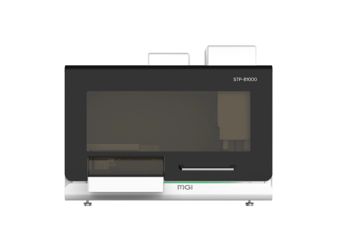
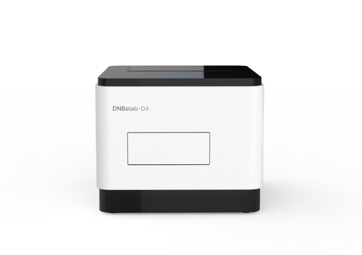
DCS Lab
DCS Lab is the first laboratory empowerment program targeting the world's cutting-edge scientific research fields. It aims to help facilitate large-scale leading multi-omics laboratories and spur critical scientific research.
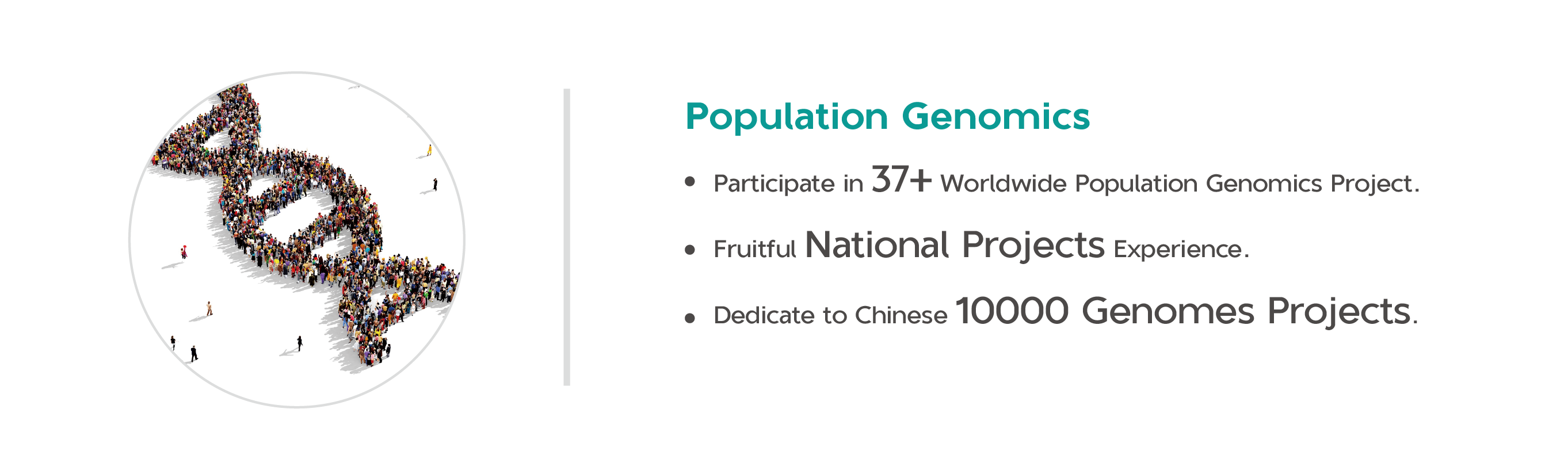
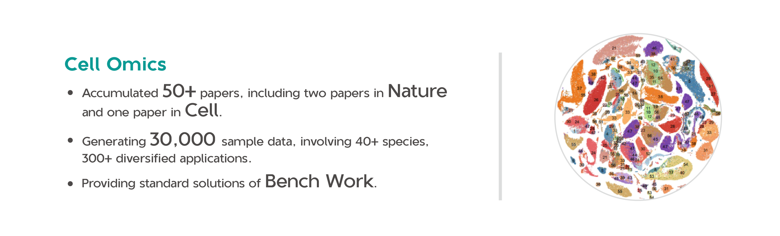
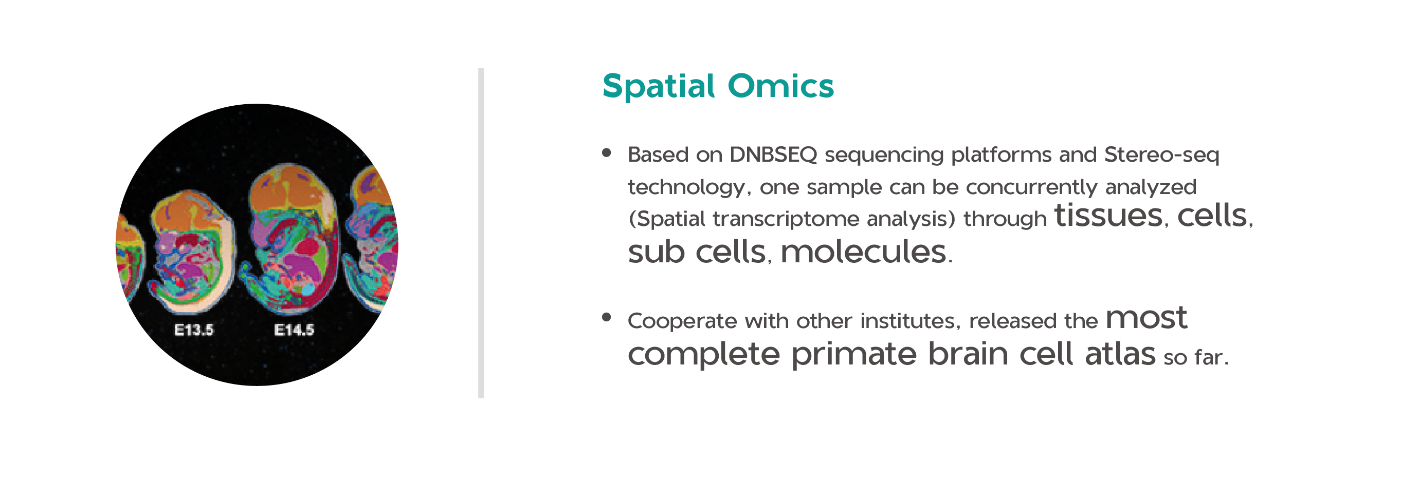
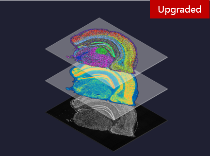
Advanced life science tools can promote scientific discoveries and research results. DCS Lab will promote the industry's development in large population genomics, single cell omics, and spatiotemporal omics through three key frontier areas (which respectively correspond to "DNA Sequencers", "Cell Omics", and "Spatial Omics" in "DCS Lab"), accelerating the scientific research empowered by emerging technologies.